CCMA - Week 13 - Day 1
Stepful・2 minutes read
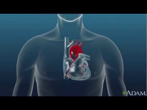
Understanding EKG basics, including sinoatrial and atrioventricular nodes, waveform characteristics, lead placement, and heart rate calculations, is crucial in diagnosing cardiac conditions. Various rhythms like sinus tachycardia, atrial fibrillation, and ventricular fibrillation present different risks, emphasizing the importance of prompt medical intervention to avoid severe complications and ensure proper heart function.
Insights
- EKG readings are crucial for understanding heart activity, with normal sinus rhythm originating from the sinoatrial node and the atrioventricular node acting as a backup.
- Differentiating between various EKG patterns, such as sinus tachycardia, atrial fibrillation, and ventricular fibrillation, is essential for diagnosing and managing cardiac conditions effectively, as each pattern presents unique risks and implications that can be life-threatening if left untreated.
Get key ideas from YouTube videos. It’s free
Recent questions
What are the key components of a normal EKG waveform?
The normal EKG waveform consists of a P-wave, representing atrial depolarization, a QRS complex indicating ventricular depolarization, and a T-wave signifying relaxation.
How can EKG artifacts affect readings?
EKG artifacts like AC interference, somatic tremor, wandering baseline, and interrupted baseline can distort readings, leading to inaccurate interpretations of heart activity.
What distinguishes sinus tachycardia from sinus bradycardia?
Sinus tachycardia is characterized by a heart rate over 100 beats per minute with a p-wave before the QRS complex, while sinus bradycardia has a slower heart rate with different waveform characteristics.
What is the difference between ventricular tachycardia and ventricular fibrillation?
Ventricular tachycardia is identified by rapid ventricular activity with no p-wave and inverted QRS complexes, while ventricular fibrillation displays chaotic electrical activity with no organized rhythm.
How does asystole differ from ventricular fibrillation?
Asystole indicates a complete stop in heart activity, while ventricular fibrillation shows unorganized electrical activity with blood pooling in the ventricles, posing life-threatening risks.
Related videos


Summary
00:00
Understanding EKG: Building Blocks and Rhythms
- EKG review: Understanding the building blocks of EKG, focusing on triggers in the heart - the sinoatrial node and the atrioventricular node.
- Normal sinus rhythm originates from the sinoatrial node, with the atrioventricular node as a backup.
- Differentiating between arrhythmias and ventricular rhythms based on waveform characteristics.
- Heart rate is beats per minute, while heart rhythm refers to the chambers' communication.
- Lead placement: Precordial chest leads (6) and limb leads (4 on extremities), ensuring symmetry in placement.
- Providers can monitor EKG readings in real-time for abnormalities and lead adjustments.
- Identifying a normal EKG waveform: P-wave signifies atrial depolarization, QRS complex represents ventricular depolarization, and T-wave indicates relaxation.
- Addressing artifacts in EKG readings: AC interference, somatic tremor, wandering baseline, and interrupted baseline.
- Calculating heart rates using the 1500 method for regular rhythms and the 6-second method for irregular rhythms.
- Differentiating between normal sinus rhythm and abnormal rhythms like sinus bradycardia based on waveform characteristics and heart rate calculations.
16:46
Cardiac EKG Patterns: Identification and Implications
- Sinus tachycardia is identified by the presence of a p-wave before the QRS complex, with a heart rate over 100 beats per minute.
- Sinus arrest, lasting longer than six seconds, indicates a pause in the heart rhythm due to the failure of the sinoatrial node and atrioventricular node.
- Atrial flutter is characterized by rapid atrial contractions that the ventricles cannot keep up with, leading to a faster heart rate but less effective activity.
- Atrial fibrillation presents as irregular sawtooth waves on the EKG, with no distinct p-wave, potentially causing blood clots due to stagnant blood flow.
- Ventricular tachycardia is recognized by the absence of a p-wave and inverted QRS complexes, indicating rapid ventricular activity over 100 beats per minute.
- Ventricular bradycardia, with wider QRS complexes and longer gaps between beats, signifies slower ventricular activity.
- Ventricular fibrillation displays chaotic electrical activity with no identifiable p-wave or organized rhythm, posing a severe risk due to ineffective heart contractions.
- In ventricular fibrillation, blood pools in the ventricles, leading to a life-threatening situation where the heart is unable to pump effectively.
- The absence of QRS complexes in ventricular fibrillation indicates a lack of organized heart activity, differentiating it from asystole where the heart has completely stopped.
- Understanding the distinctions between various EKG patterns is crucial in diagnosing and managing different cardiac conditions, each presenting unique risks and implications.
33:56
Heart irregularities and patient intake scenarios discussed.
- The heart's ventricles are trying to push blood through, but there is an issue with communication from the Sinoatrial node.
- Asystole indicates the heart has stopped completely, while in Vfib, part of the heart is still contracting but not effectively communicating.
- Vfib leads to unorganized electrical activity, causing blood to pool in the heart's ventricles.
- These heart irregularities are serious and can lead to death if untreated, potentially causing blood clots and heart shutdown.
- The speaker's daughter experienced heart issues at birth due to missing arteries and valves, leading to blood pooling in the heart.
- Prompt medical intervention saved the speaker's daughter, who self-corrected her heart issue while in transit to the hospital.
- The heart compensates for deficiencies but cannot sustain them long-term, eventually leading to heart failure.
- The group project focuses on patient intake and interview scenarios, with members role-playing different roles and responsibilities.
- The project involves reviewing patient intake forms, determining the relevance of medical reports to the patient's condition, and practicing role-play scenarios.
- The lesson progresses to medical office administration, covering topics like written and telephone communication, electronic health records, and medical insurance and billing.
50:34
"Role-playing scenarios and group projects for graduation"
- The scenarios of role-playing as a patient for the intake interview and Miss Jackson are unrelated, with each requiring separate responses and actions.
- Completing both scenarios is mandatory for graduation, with group projects being essential for completion. If any group project is missing, completing the written submission will provide 50% credit, although full participation is required for full credit. This week is ideal for catching up on any missing assignments.


