Neuroanatomy | Anatomy | FARRE 2.0 | MBBS Prof 1 | Dr. Pradeep
PW MedEd・63 minutes read
Neuroanatomy lecture delves into brain structures like sulci and gyri, Broadman's classification, specific areas for sensory and motor functions, blood supply details, and key brain structures, culminating in an encouragement for questions with expressions of gratitude.
Insights
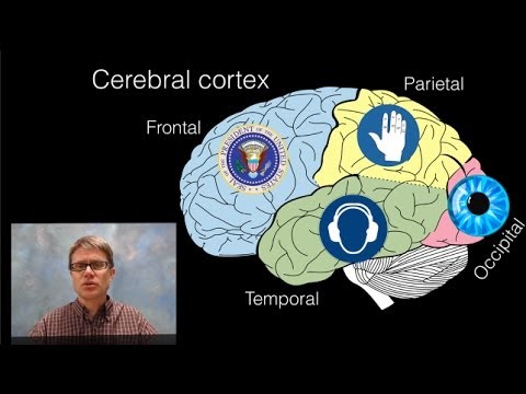
- Brain areas are classified by Broadman into primary areas for sensation perception and association areas for interpretation, with specific regions like 31-32 for sensory functions and 5 and 7 for sensory association, highlighting the brain's specialized functions.
- Blood supply in the brain is intricate, with the middle cerebral artery providing for motor and sensory areas, the posterior cerebral artery for the visual cortex, and the macular supply by the middle cerebral artery, emphasizing the importance of vascular support in brain function.
- Understanding the Venus system of the brain, the intricate network of veins and arteries, is crucial for maintaining proper blood flow and drainage, ensuring the brain's optimal functioning and health.
Get key ideas from YouTube videos. It’s free
Recent questions
What are the primary areas of the brain responsible for perception?
Areas 31-32 for sensory functions, 5 and 7 for sensory association.
Which brain areas are involved in visual processing?
Occipital lobe areas 17, 18, and 19 for visual processing.
What is the role of Broca's area in the brain?
Broca's area (areas 44 and 45) is responsible for motor speech functions.
What arteries supply the motor and sensory areas of the brain?
Middle cerebral artery for motor and sensory areas, posterior cerebral artery for visual cortex.
What structures are key in the brain's venous system?
Superior cerebral vein, inferior cerebral vein, anterior cerebral vein, middle cerebral vein.
Related videos


Summary
00:00
Brain Superolateral Surface: Anatomy and Functions
- Neuroanatomy lecture focusing on the superolateral surface of the brain
- Discussion on sulci and gyri of the brain's superolateral surface
- Broadman's classification of brain areas into primary and association areas
- Primary areas for perception of sensations and association areas for interpretation
- Specific brain areas like 31-32 for sensory functions, 5 and 7 for sensory association
- Occipital lobe areas 17, 18, and 19 for visual processing and association
- Temporal lobe areas 41 and 42 for auditory functions, including sensory speech area 22
- Frontal lobe areas 44 and 45, known as Broca's area, for motor speech functions
- Discussion on corticonuclear and corticospinal fibers originating from area 4
- Blood supply details, including middle cerebral artery for motor and sensory areas, and posterior cerebral artery for visual cortex, with a special mention of macular supply by middle cerebral artery.
18:53
Cerebral Arteries and Veins: An Overview
- The anterior cerebral artery is a continuation of the internal carotid artery, while the middle cerebral artery is a continuation of the internal carotid artery.
- The vertebral arteries join to form the basilar artery, which then divides into terminal branches, including the posterior cerebral artery.
- The vertebral artery is a branch of the first part of the subclavian artery.
- The Circle of Willis is formed by the anterior communicating artery, anterior cerebral artery, internal carotid artery, posterior communicating artery, and posterior cerebral artery.
- The optic nerve, optic chiasm, optic tract, and mammillary bodies are key structures in the brain.
- The medulla contains pyramids, olives, and the inferior cerebellar peduncle.
- The veins of the brain include the superior cerebral vein, inferior cerebral vein, anterior cerebral vein, and middle cerebral vein.
- The basil vein of Rosenthal is formed by the anterior cerebral vein, deep middle cerebral vein, and striate veins.
- The internal cerebral veins are formed by the thalamic vein, choroidal vein, and septal vein, which then join to form the great cerebral vein of Galen.
- Surface anatomy of the brain includes the thalamus, caudate nucleus, thalamic vein, choroidal plexus, and septal vein.
37:25
Brain Anatomy: Understanding Venus System and More
- Understanding the Venus system of the brain involves the flow of blood from the brain's supply to the Venus drainage.
- Diagrams are crucial for explaining brain anatomy, with detailed labeling enhancing understanding and potentially leading to better marks in exams.
- The corpus callosum consists of the rostrum, genu, trunk, and splenium, with commissural fibers connecting similar areas in opposite hemispheres.
- The septum pellucidum acts as a partition between the corpus callosum and the fornix, leading to the lateral ventricle.
- The lamina terminalis is located near the optic chiasm, with structures like the pineal gland and habenular commissure forming the epithalamus.
- The diencephalon comprises the thalamus, hypothalamus, epithalamus, metathalamus, and subthalamus, with the lateral ventricle being part of the diencephalon.
- The subthalamic nucleus is part of the basal ganglia, along with the caudate nucleus, putamen, globus pallidus, and substantia nigra.
- The hypothalamic sulcus divides the diencephalon into the thalamus and hypothalamus, with the third ventricle being the cavity between them.
- The third ventricle's roof is formed by the fornix, floor by the optic chiasm, pituitary stalk, and mamillary bodies, and walls by various structures like the thalamus and hypothalamus.
- Communication between the lateral ventricle and the third ventricle occurs through the foramen of Monro, while the aqueduct of Sylvius connects the third ventricle to the fourth ventricle.
53:46
Cranial Nerves and Neuroanatomy Essentials
- Facial cecus is formed by fibers of the facial nerve winding around the abducent nucleus, creating an elevation outside.
- The hypoglossal triangle and vagal triangle are correlative structures, with the hypoglossal triangle housing the hypoglossal nucleus and the vagal triangle containing the dorsal nucleus of the vagus.
- Important topics for neuroanatomy include the brain stem, midbrain, pons, medulla, pyramids, olives, and cerebellar peduncles.
- Essential cranial nerves to know are the 7th, 9th, and 12th, with functional components, nucleus names, and attachment to the brain stem crucial for understanding.
- The third nerve emerges from the ventral aspect of the midbrain, while the fourth nerve emerges from the dorsal aspect, with nuclei located at different levels in the midbrain.
- The fifth nerve is attached at the junction of the pons with the middle cerebellar peduncle, and the sixth, seventh, and eighth nerves are located at the ponto-medullary junction.
- Nerves between the olives and the inferior cerebellar peduncle include the 9th, 10th, and cranial accessory nerves, while the 12th nerve is between the pyramids and olives.
- The third, fourth, and sixth nerves exit through the foramen to reach the orbit, passing through the middle cranial fossa and related to the cavernous sinus.
- The glossopharyngeal nerve supplies the stylopharyngeus muscle, parotid gland, and taste sensations to the posterior 1/3 of the tongue, with functional components including special visceral efferent and general visceral efferent.
- The nucleus ambiguous is crucial for supplying the stylopharyngeus muscle, while the inferior salivatory nucleus is involved in supplying the parotid gland, with the nucleus of the tractus solitarius responsible for carrying special sensations from the tongue.
01:10:23
"Glossopharyngeal nerve and neuroanatomy essentials"
- Glossopharyngeal nerve exits through the jugular foramen outside the skull.
- Passes between superior and middle constrictor muscles to reach the stylopharyngeus muscle.
- Glossopharyngeal nerve then deepens to the hyoglossus muscle to supply the tongue.
- Neuroanatomy exams focus on cardinal structures like caudate nucleus, thalamus, and lentiform nucleus.
- Internal capsule contains projection fibers, with anterior limb between caudate and lentiform nuclei.
- Posterior limb lies between thalamus and lentiform nucleus.
- Retrolentiform part carries visual pathway fibers, while sublentiform part carries auditory pathway fibers.
- Corticonuclear fibers pass through genu, corticospinal fibers through posterior limb of internal capsule.
- Climbing fibers stimulate Purkinje cells directly, while mossy fibers stimulate granule cells first.
- Cerebellar circuit involves climbing and mossy fibers, with Purkinje cells inhibiting deep cerebellar and lateral vestibular nuclei.
01:29:15
"Grateful farewell with encouragement for questions"
- Encouragement to reach out for any questions or uncertainties
- Expressing gratitude for the audience's patience and attention
- Signing off with thanks and goodbye


