The Skeletal System: The Appendicular Skeleton - Ch. 8
idiobrett・28 minutes read
Chapter 8 discusses the anatomy of the appendicular skeleton, focusing on bones of the upper and lower limbs, shoulder girdles, and pelvis, highlighting differences between male and female anatomy, particularly in relation to childbirth and pelvic structure. The development of limb girdles and limbs from mesoderm between weeks four to eight after fertilization plays a crucial role in forming the upper and lower limbs, with significant growth occurring during this period, leading to the development of distinct features like hands, feet, and facial structures.
Insights
- The female pelvis is wider and shallower compared to the male pelvis, impacting childbirth due to anatomical differences in size and structure.
- The limb girdles and limbs develop from mesoderm between weeks four and eight post-fertilization, with significant growth and formation occurring during this period, particularly in the upper and lower limbs, leading to the gradual formation of distinct human features like hands, feet, eyes, and facial structures.
Get key ideas from YouTube videos. It’s free
Recent questions
What are the bones of the pectoral girdle?
Clavicle and scapula
Related videos
TeachMeAandP
The skeletal system: Appendicular Skeleton

RegisteredNurseRN
Regional Terms Anatomy - Body Parts Name | Nursing Medical Terminology Made Easy

Dr. Rakshita Singh
Skeletal System with 3D Model- Locomotion and Movement | NEET 2024- Class 11

Kto12 Lessons
Science 6 Quarter 2 Module 1 Lesson 1 - The Skeletal System

CrashCourse
Joints: Crash Course Anatomy & Physiology #20


Summary
00:00
Appendicular Skeleton: Limbs, Girdles, and Joints
- Chapter 8 begins on page 242, focusing on the appendicular skeleton involved in body movements.
- The appendicular skeleton includes bones of the upper and lower limbs, along with the shoulder girdles.
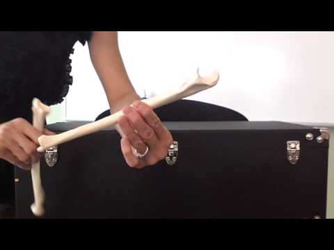
- The pectoral girdle consists of the clavicle (collarbone) and scapula (shoulder blade).
- The scapula's glenoid cavity attaches to the humerus, forming the shoulder joint.
- The humerus articulates with the scapula proximally and with the radius and ulna distally.
- The ulna and radius form the forearm, allowing for rotation.
- Carpal bones are arranged in two rows, proximal and distal, articulating with the radius and ulna.
- Metacarpals make up the palm and back of the hand, articulating with phalanges (fingers).
- The pelvis consists of two hip bones (coxal bones) articulating with the sacrum.
- The male and female pelvis differ in size and structure, with the female pelvis wider and shallower for childbirth.
16:29
Female Pelvic Anatomy and Childbirth Considerations
- Female anatomy is shaped differently due to anatomical and physiological reasons, such as wider pelvis for childbirth.
- The pubic arch is wider in females than in males, affecting their center of gravity.
- Lateral views of the male and female pelvis show differences in curvature and width.
- The pelvic outlet is wider in females and narrower in males, impacting childbirth.
- Doctors measure the pelvic axis during pregnancy to determine if the baby's head can fit through the birth canal.
- The pelvis protects reproductive organs, with females having internal organs within the pelvis.
- The femur is the longest, heaviest, and strongest bone in the body, articulating with the hip and tibia.
- The patella articulates with the femur and is held in place by a tendon.
- The lower leg consists of the tibia and fibula, with the tibia articulating with the femur and tibia tuberosity.
- The foot has two arches, longitudinal and transverse, supported by ligaments and tendons for weight distribution and leverage while walking.
32:00
Embryonic Limb Development: Weeks Four to Eight
- The skeleton of the limb girdles and limbs develops from mesoderm between week four and week eight after fertilization, with significant growth and formation occurring during this period, particularly in the upper and lower limbs.
- By about six weeks, the hand and foot start to take shape, along with the development of the eye and orbitals, while the pharyngeal arches continue to progress, leading to a more defined appearance resembling a person as the weeks progress.


