Layers of the heart | Human anatomy and physiology | Health & Medicine | Khan Academy
khanacademymedicine・2 minutes read
Blood flows through the heart from atrium to ventricle, lungs, and finally back to the heart, with atrioventricular valves crucial for maintaining direction. The heart's structure includes three layers - endocardium, myocardium, and pericardium - each with specific functions in supporting heart function and circulation.
Insights
- Atrioventricular valves like the tricuspid and mitral valves are crucial in ensuring blood flows in the correct direction through the heart, preventing backflow that could disrupt circulation.
- The heart's structure, including the three layers of heart muscle (endocardium, myocardium, pericardium), the role of the interventricular septum in maintaining separation between the ventricles, and the development of the pericardium around the growing heart, all contribute to the heart's efficient functioning and protection.
Get key ideas from YouTube videos. It’s free
Recent questions
What is the pathway of blood flow in the heart?
Blood flows from the right atrium to the right ventricle, then to the lungs, and finally to the left atrium and left ventricle.
What are the atrioventricular valves responsible for?
The atrioventricular valves, the tricuspid valve, and the mitral valve are crucial for maintaining blood flow direction in the heart.
How are the atrioventricular valves prevented from flipping back?
The atrioventricular valves are tethered to prevent flipping back, with papillary muscles and chordae tendineae playing a key role in keeping them in place.
What can happen if a ventricle is too strong in the heart?
If a ventricle is too strong and breaks a cord, the valve may flip back, causing blood to flow in the wrong direction.
What are the layers of the heart muscle?
The heart muscle has three layers: endocardium, myocardium, and pericardium (with two layers, visceral and parietal). The endocardium is thin and similar to blood vessel linings, while the myocardium is where contractile muscle and energy use are concentrated. The pericardium has an inner visceral layer (epicardium) hugging the heart and an outer parietal layer, with a gap between them containing fluid.
Related videos
Dr. John Campbell
Cardiovascular System 1, Heart, Structure and Function

CrashCourse
The Heart, Part 1 - Under Pressure: Crash Course Anatomy & Physiology #25

OCC Anatomy
Heart 1
khanacademymedicine
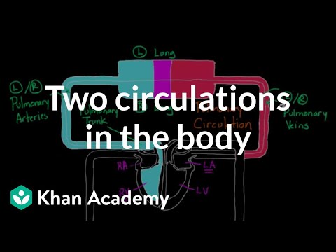
Two Circulations in the Body | Circulatory system physiology | NCLEX-RN | Khan Academy

khanacademymedicine
Flow through the heart | Circulatory system physiology | NCLEX-RN | Khan Academy


Summary
00:00
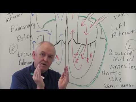
Anatomy of the Heart: Blood Flow and Structure
- Blood flows from the right atrium to the right ventricle, then to the lungs, and finally to the left atrium and left ventricle.
- The atrioventricular valves, the tricuspid valve, and the mitral valve are crucial for maintaining blood flow direction.
- The atrioventricular valves are tethered to prevent flipping back, with papillary muscles and chordae tendineae playing a key role.
- If a ventricle is too strong and breaks a cord, the valve may flip back, causing blood to flow in the wrong direction.
- The interventricular septum has a membranous and muscular part, with VSD (ventricular septal defect) common in the membranous part.
- The heart muscle has three layers: endocardium, myocardium, and pericardium (with two layers, visceral and parietal).
- The endocardium is thin and similar to blood vessel linings, while the myocardium is where contractile muscle and energy use are concentrated.
- The pericardium has an inner visceral layer (epicardium) hugging the heart and an outer parietal layer, with a gap between them containing fluid.
- The pericardium forms around the growing heart in a fetus, enveloping it and eventually folding back on itself to create the layers seen in the adult heart.


