Cardiovascular | Structures and Layers of the Heart
Ninja Nerd・21 minutes read
The heart is a vital organ located in the mediastinum, with a weight of 200 to 300 grams and various chambers receiving and pumping blood. Valves, chordae tendineae, and different arteries play crucial roles in the heart's structure and function.
Insights
- The heart is located in the mediastinum, two-thirds to the left of the mid-sternal line, weighing around 200 to 300 grams, and is roughly the size of a fist.
- The heart's structure includes chambers like the atria, receiving blood from specific veins, with valves separating the atria from the ventricles, and chordae tendineae anchoring the valves to prevent backflow.
Get key ideas from YouTube videos. It’s free
Recent questions
Where is the heart located in the body?
In the mediastinum within the thoracic cavity.
What are the chambers of the heart called?
Atria and ventricles.
What are the functions of heart valves?
Prevent backflow and regulate blood flow.
How does the heart pump blood to the body?
Through the pulmonary and aortic semilunar valves.
What are the layers of the heart?
Endocardium, myocardium, and epicardium.
Related videos
Dr. John Campbell
Cardiovascular System 1, Heart, Structure and Function

Peekaboo Kidz
How Your Heart Works? - The Dr. Binocs Show | Best Learning Videos For Kids | Peekaboo Kidz

BTEC Applied Science Help
BTEC Applied Science: Unit 5 B1 The Heart

CrashCourse
The Heart, Part 1 - Under Pressure: Crash Course Anatomy & Physiology #25
khanacademymedicine
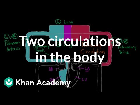
Two Circulations in the Body | Circulatory system physiology | NCLEX-RN | Khan Academy


Summary
00:00
Anatomy of the Heart: Key Points
- The heart is situated in the mediastinum within the thoracic cavity, shifted two-thirds to the left of the mid-sternal line.
- The apex of the heart points towards the left hip, while the base points towards the right shoulder.
- The heart weighs approximately 200 to 300 grams, about the size of a fist.
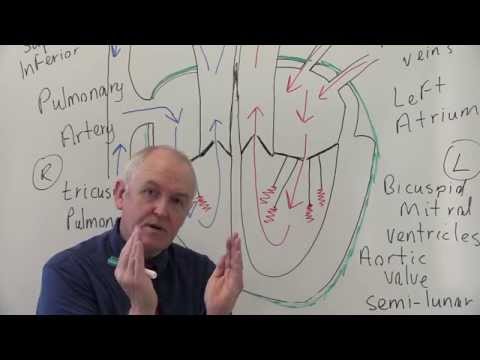
- The heart consists of chambers, with the top chambers known as atria, such as the right atrium and left atrium.
- The right atrium receives blood from the inferior vena cava, superior vena cava, and coronary sinus.
- The left atrium receives blood from the pulmonary veins, bringing oxygenated blood from the lungs.
- Valves separate the atria from the ventricles, including the tricuspid valve on the right side and the bicuspid or mitral valve on the left side.
- The valves are made up of three layers - zona spongy, zona fibrosis, and zona ventricular, with an endothelial lining.
- Chordae tendineae are collagen cords that anchor the valves to papillary muscles, preventing backflow.
- The right ventricle pumps blood to the pulmonary trunk through the pulmonary semilunar valve, while the left ventricle pumps blood to the aorta through the aortic semilunar valve.
13:30
Anatomy of the Heart and Major Arteries
- The right pulmonary artery leads to the right lung.
- The ascending aorta separates the left ventricle from the aorta.
- The aortic arch branches into the brachiocephalic, left common carotid, and left subclavian arteries.
- The heart layers consist of the endocardium, myocardium, and epicardium.
- The epicardium can also be referred to as the visceral layer of the serous pericardium.
- The fibrous pericardium anchors, protects, and prevents the heart from overfilling with blood.


